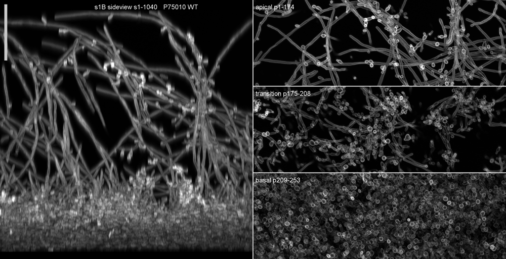
Our overall interest is in understanding the biophysical principles by which cells self-organize and carry out essential mechanical functions, and in working out measurement methods to test the relevant hypotheses. Current projects include (1) measurement of hyphal growth in fungal cells, and how this is related to invasion and virulence in Candida albicans, and (2) development of high-resolution imaging methods for large-scale objects such as biofilms, microbial colonies, embryos, organoids, and thick tissue sections. Most of my work is done with C. albicans in collaboration with Aaron P. Mitchell (Univ. Georgia) and Joel McManus (CMU). We have developed refractive index matching methods for fast, high-quality clarification of fixed C. albicans colonies and biofilms. Recently, we extended our clarification methods to living biofilms by use of iodixanol, a non-ionic, water-soluble, organo-iodine compound with moderately high specific refractivity that is used medically as an X-ray contrast agent. In 2024, our optical innovations resulted in a US patent on a microscope objective design that provides a basis for development of a low-cost instrument for fast 3D imaging of clarified specimens.

