High-resolution spatiotemporal organization of the T cell membrane
The membrane plays a pivotal role as the primary platform for T cell activation and function. During initial contact, T cell membrane protrusions scan the surface of antigen-presenting cells (APC) in search of antigens. The engagement of the T cell receptor (TCR) with its specific antigen triggers T cell activation, leading to the formation of the immunological synapse, a tight cell-cell junction with the APC. This process involves a drastic reorganization of the T cell cytoskeleton, plasma membrane, surface receptors, and intracellular signaling molecules. Within the immunological synapse, TCRs recruit other surface receptors, along with kinases and adaptor molecules, to form microclusters that serve as fundamental signaling units. Therefore, the spatiotemporal organization of the T cell membrane plays a crucial role in T cell signaling and sensitivity.
We aim to determine the nanoscale membrane organization and dynamics of these membrane proteins and understand how this nanoscale organization and dynamics contribute to T cell activation and inhibition.
a. T cell membrane on the nanoscale
We use Expansion Microscopy (ExM) to achieve nanoscale resolution of the T cell in all three dimensions. Expansion microscopy (ExM) is an advanced method for super-resolution imaging that uses a chemical process to preserve and uniformly expand the specimen, enabling resolution capabilities as fine as 50 nm. The pictures on the top panels (provided by Ellie Lai) of the figure presented below show examples of pre-expansion and post-expansion images of T cells, stained with the membrane marker and microvilli marker. The comparison of these images before and after expansion demonstrates the significant enhancement in 3D spatial resolution achieved through ExM. This technique reveals nanoscale membrane topology and distinct nanoclusters of membrane receptors prior to antigen engagement.
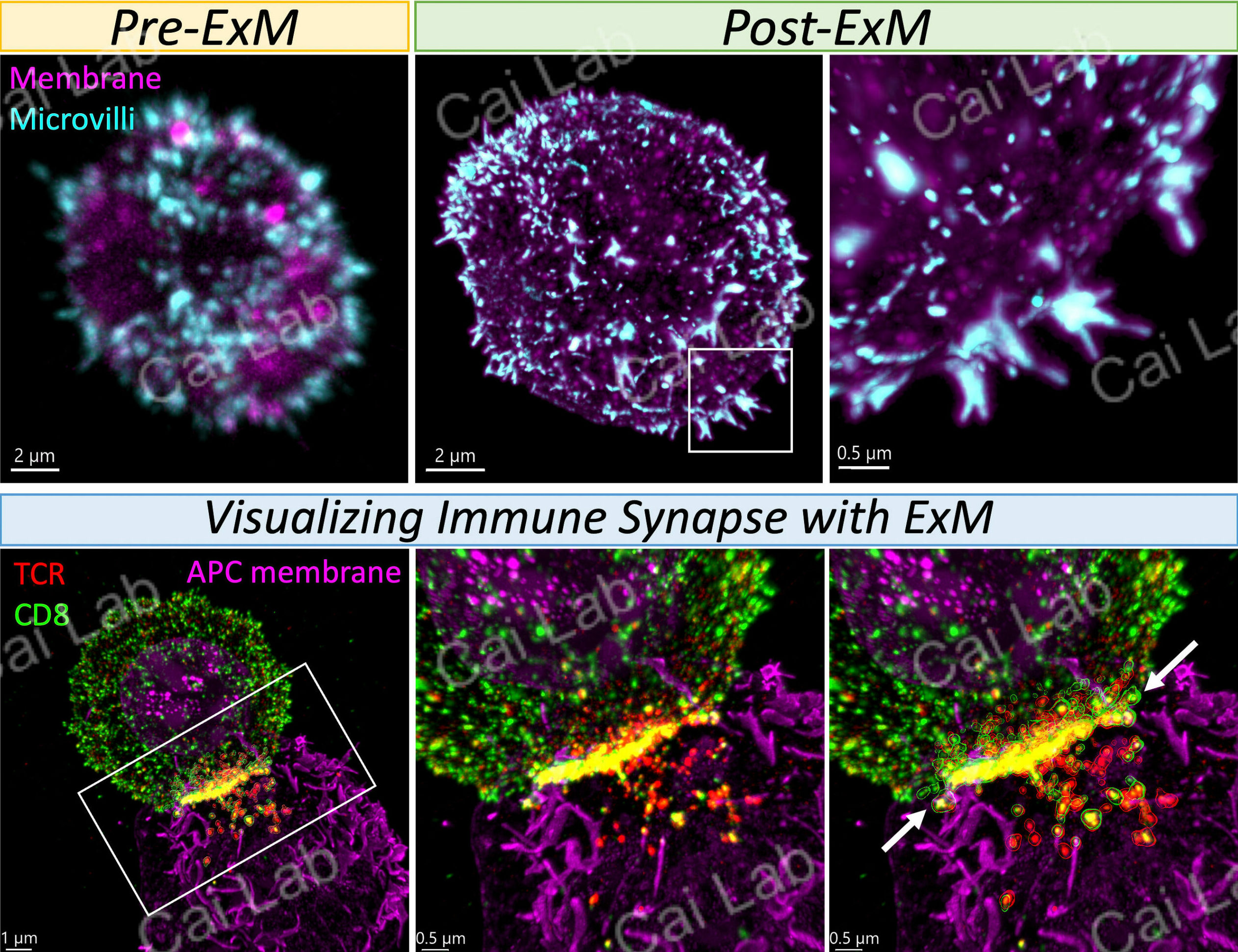
Next, we imaged the organization of the cell membrane together with receptors in the immunological synapse formed between T cells and APCs. To achieve high-resolution images, we expanded the conjugated T cell: APC complex. These high-resolution images enabled us to reconstruct the cell surface and facilitated the visualization of membrane topology at the contact sites, shown on the bottom panels of the above figure (provided by Suzi Kim).
b. Roles of T cell membrane organization in T cell signaling
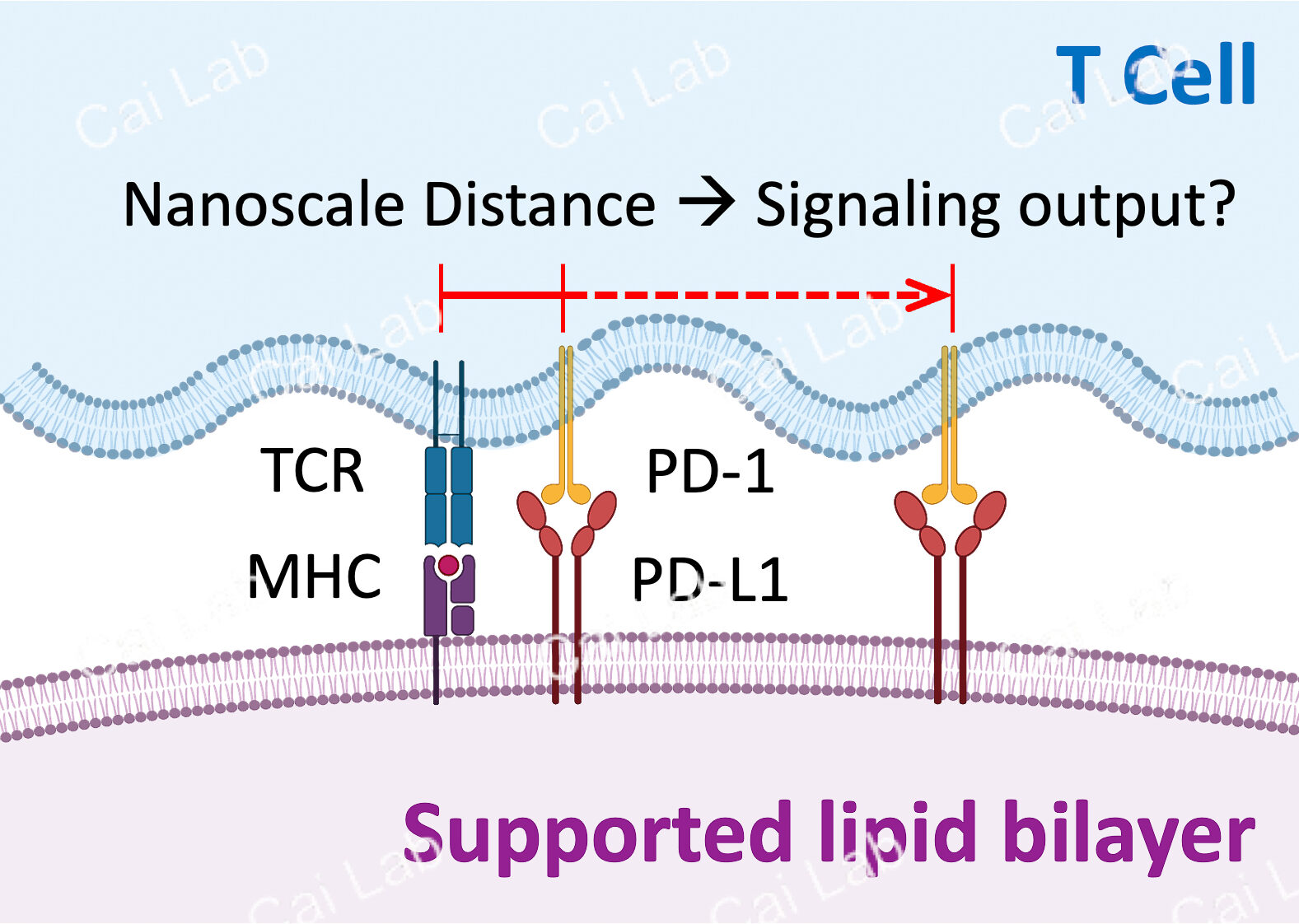
The main goal of this project is to investigate how receptor organization influences T cell signaling. Our specific focus lies on examining the interaction between the T cell receptor (TCR) and the inhibitory receptor programmed cell death protein 1 (PD-1). Previous studies have indicated that PD-1 often co-localizes with TCR microclusters and exerts inhibitory effects on TCR signaling when in close proximity. In this project, we utilize supported lipid bilayers to analyze the signaling output in relation to the nanoscale distance between TCR and PD-1, as depicted in the illustration below (provided by Hamid Akhbariyoon).
_______________________________________________________________________________________
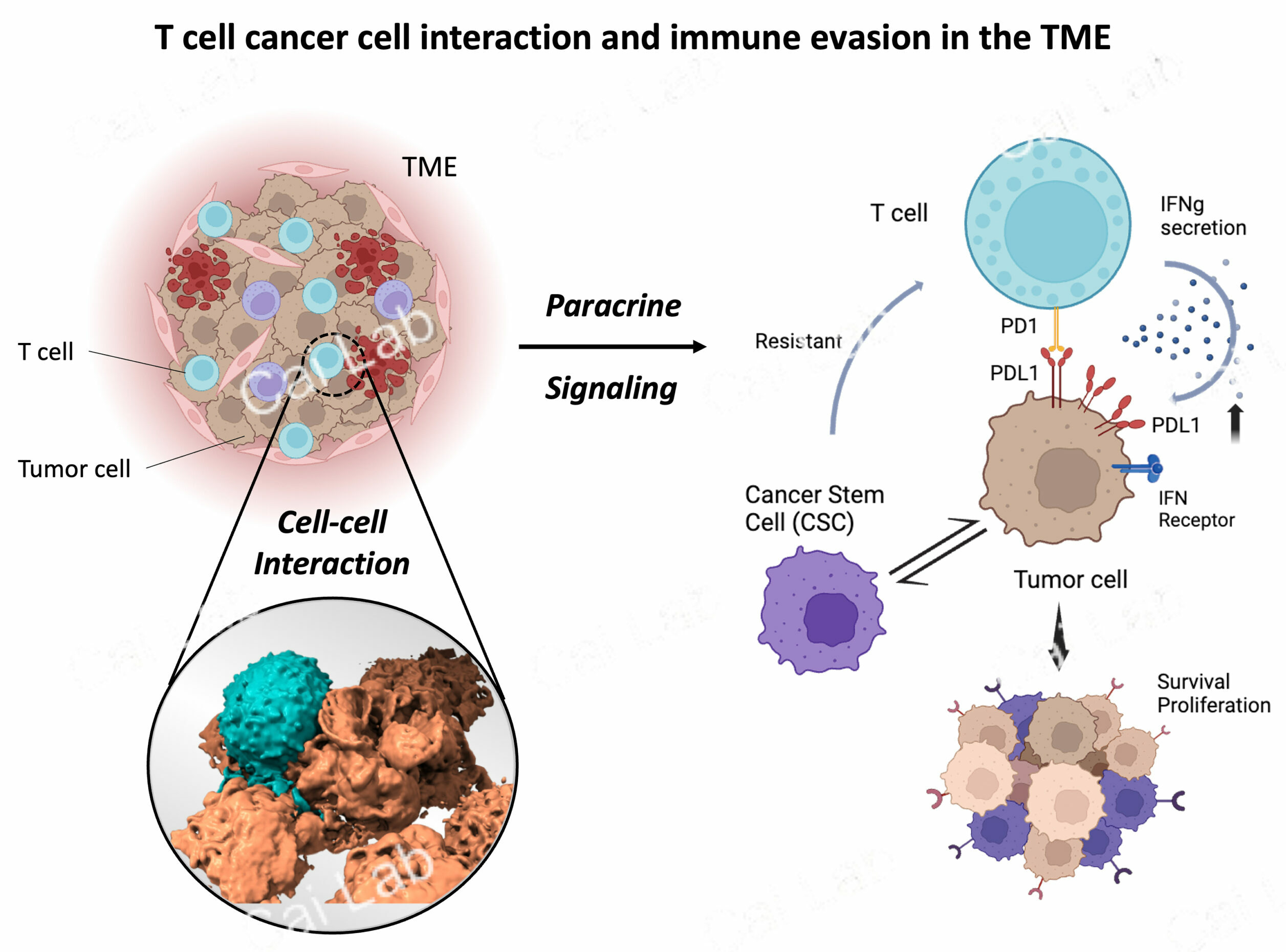
Cancer cell and T cell interactions in the tumor microenvironment and their roles in immune evasion
In the tumor microenvironment (TME), cancer cells have developed strategies to evade immune cytotoxicity. It is crucial to understand the communication and interactions between cancer cells and T cells in this microenvironment. One way of communication is through direct cell-cell contact. In this context, we explore how T cells navigate the TME, survey cell surfaces to recognize antigens, and examine how the interaction between T cells and cancer cells may lead to cell-cell contacts or dysfunctional synapses, if present. Another way of cross-talk between cancer cells and T cells is through paracrine signaling. Our specific interest lies in the mechanism of IFN-γ’s function in increasing expression of PD-L1 and increasing level of cancer cell stemness which contribute to cancer cell immune evasion. (Image provided by En Cai and Yue Zhang)