My lab asks how the brain transforms thought into action. To do so, we apply advanced computational methods to the neural dynamics during tasks tailored to answer our specific mechanistic questions. Where no tool exists, we take the initiative to pioneer our own – then release it as a user-accessible package with the hopes of enabling broader use and deeper scientific impact. This approach has created a highly collaborative and engaging environment within the lab, across the university, and internationally. Our scientific vision is anchored by two interlocking themes: (1) dissecting the precise computational roles of basal ganglia circuit elements in movement and decision-making, and (2) building and deploying open-source behavioral quantification tools that enable rigorous, unbiased analysis of naturalistic behavior at scale. In both cases, we seek to apply these circuit and computational frameworks to address clinically meaningful questions in movement disorders, psychiatric disease, and chronic pain.
Theme 1: Basal Ganglia Circuit Computations and Movement Control
The central goal of our research program is to understand how the basal ganglia and its neural partners control movement and behavioral decisions. Much of this work has focused on establishing the specific computational function of the opponent pathways of the basal ganglia – the direct and indirect pathway neurons of the striatum. The prevailing theory holds that these pathways respectively encode some form of facilitation (“go”) and suppression (“no-go”) signals that select for particular actions. However, other results suggested a role in learning or motor control. Through targeted experimental manipulations, we established that increasing the relative activity of either pathway will shape movement speed in a manner that is specific to the policy that produced the activity change, without any ‘action selection’ effects on motor output (Yttri and Dudman, Nature; Yttri and Dudman, Movement Disorders; Fig 1). These findings imply that the populations act like an adaptive volume knob on headphones, learning how to adjust to each environment – but without changing the song.
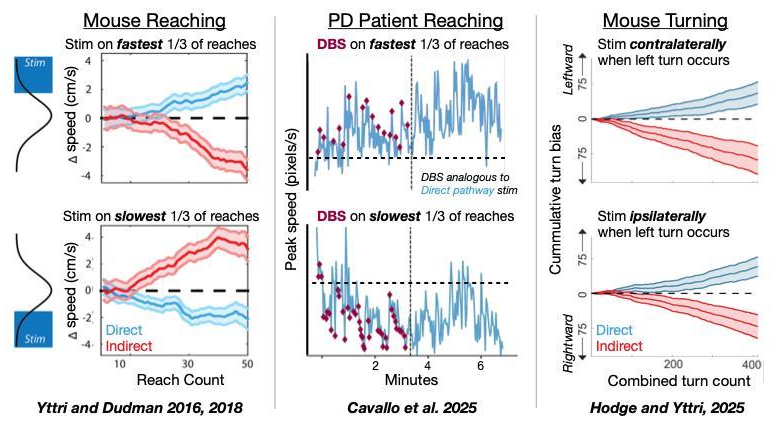
Fig 1: Consistent, pathway-specific effects across different behaviors and species.
We next established that these findings are broadly applicable to striatal function, not just a rewarded, over-trained task. To determine if policy-based reinforcement learning is a core tenant of basal ganglia function, we expanded into wider contexts. First, we developed task-free paradigms that were specifically designed to directly dissociate action-based and reinforcement-based accounts of basal ganglia activity, e.g. if direct pathway neurons are stimulated in a physiologically-relevant manner whenever an animal stops walking, will it be invigorated to move or reinforced to remain still (Hodge and Yttri, Cell Reports; Badyna et al., In Review; Fig 1). In every case, the results were only consistent with the reinforcement hypothesis. These results demonstrate that striatal pathway activity does not encode a fixed action label but instead reinforces along whatever policy parameter is active at the time of stimulation, providing strong causal evidence for the policy-based control framework.
We then substantially extended and sharpened this framework with a theoretical piece suggesting that a policy-based RL model best accounts for both basal ganglia biology and behavior (Hodge et al., Current Opinion: Behavioral Sciences). The dominant action value encoding framework breaks down badly when removed from reductionist experimental contexts and instead is faced with real-world behavioral complexity. By connecting observations from computational theory, biological cytoarchitecture, and neural dynamics, this Perspective argues that the fundamental computation of this circuit across any context is as a policy-based controller.
To complete this picture, we asked a complementary question: if the basal ganglia are not selecting actions, what is? By bilaterally aspirating the caudal forelimb area of motor cortex (MC) in mice trained on a joystick reaching task, we captured both the immediate and long-term neurobehavioral consequences of removing motor cortex input to striatum starting on the day of lesion (Nicholas and Yttri, Neuron). On the neural side, movement-related activity in both striatal projection neurons and fast-spiking interneurons was absent following the lesion, even during those reaches that did occur. When striatal activity eventually re-emerged in later post-lesion epochs, it no longer encoded movement vigor or kinematics – suggesting that the representation of these values was removed with the MC lesion. Together, these studies outline a circuit architecture in which MC generates and conveys the motor command to be executed, while the striatum acts downstream to modulate the kinematic parameters of that command based upon complex relationships learned in a computationally effective manner.
Psychiatric and Clinical Dimensions
As a personal highlight of my research career, we undertook the somewhat rare step of translating these basic biology ideas to the clinic. Specifically, we hypothesized that if the basal ganglia’s primary function is pushing and pulling behavioral performance according to the policies it encodes, then applying the selective stimulation paradigm from our original study with DBS should be sufficient to push patients away from the slowness of movement that defines Parkinson’s disease. After only a few brief pulses of selective stimulation, we were able to reproduce these same bidirectional reinforcement effects in several Parkinson’s patients. While I only contributed to the design, analysis and writing, I am exceedingly proud to see the years of collaborative effort published (Cavallo et al., Science Advances; Fig 1) and featured as the Keynote Lecture at the upcoming OptoDBS Conference – to be jointly delivered alongside my friend and co-author, Julian Neumann.
In our own rodent work, we also discovered the only rodent model of Freezing of Gait (FOG), a common product of Parkinson’s disease. MC-lesioned mice exhibit a profound inability to update motor state when context demanded it — freezing midstride at the threshold between two hallways in a manner strikingly reminiscent of the clinical phenotype (Nicholas and Yttri, Neuron). This occurred while retaining the ability to locomote normally in open space. We are actively pursuing funding (NIH IGNITE R01, Parkinson’s Disease Foundation) to characterize the circuit dynamics that produce pathological freezing in our model and to develop a cell-type specific, readily inducible version of the model via optogenetic inhibition.
Alongside our Parkinson’s-related work, projects within our lab have explored how basal ganglia circuits relate to processing aversion signals in psychiatric conditions (Geramita et al., Journal of Neuroscience Research). In a 2025 paper (Geramita et al., Nature Neuroscience; Fig 2), we created the first rodent experimental paradigm that reliably evokes hesitation, a pause in the face of uncertainty. Hesitation, and its inverse – impulsivity, are central to several psychiatric disorders including OCD and chronic anxiety. Coupling our experimental paradigm with cell-type specific electrophysiology, stimulation, and inhibition, we demonstrated that indirect – but not direct – pathway neurons in dorsomedial striatum mediate this slowing of transitions between behaviors. Our findings indicate that the basal ganglia circuits controlling hesitation are distinct from those controlling other forms of behavioral inhibition, such as cue-induced stopping. This work received widespread public interest – including interviews with CNN.com, KCBS News Radio, and Popular Science.
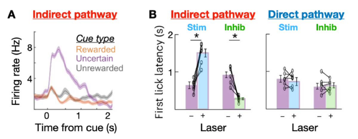
Fig 2: A) Indirect pathway neurons demonstrate robust, selective responses to ‘uncertainty cues’, tones that were predictive of potential reward but not reward outcome. B) Stimulation and inhibition of indirect, but not direct pathway neurons affected hesitation. These effects were abolished during predictive cues (not shown), indicating a strong contextual component.
Future Directions
Learning in a continuous action-reward space. As the next step to pursue the foundational computations of the basal ganglia and cortico-striatal axis, we have created a task wherein performance and reward are distributed semi-continuously throughout space (Fig 3A). Using a semicircle arena (radius = 30cm), we map a gaussian distribution (STD=12.5°) of reward probabilities over ports arranged at 5° intervals. Despite having no cues or other instructive stimuli beyond the receipt or absence of a reward, mice readily learn to perform this task. After learning the initial environment, we then shift the mean or the variance of the distribution (mean shift example in Fig 3B). Multiple shifts can be performed in the same animal. This task requires motor decisions analogous to the continuous parameters of amplitude and speed in the reaching task, but in such a way that all options have an equal cost and the experimenter has exquisite control of the decision environment.
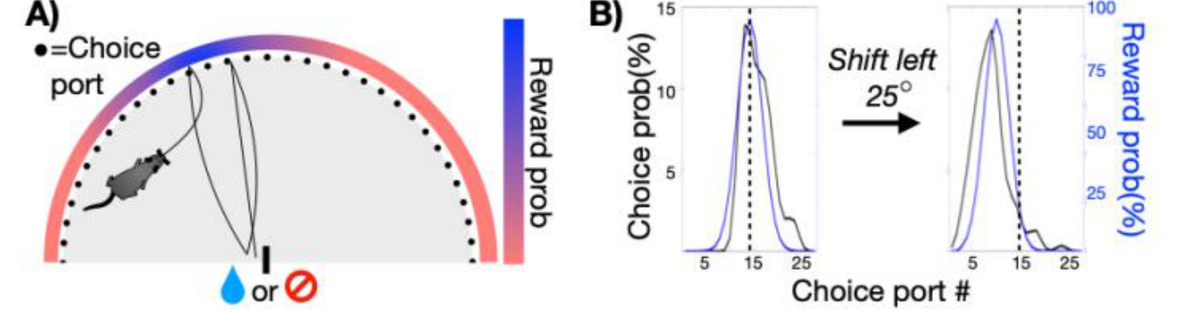
Fig 3: A) Schematic of Continuous Action/Reward Landscape (CARL) task wherein a continuous reward probability is mapped onto physical space. For each trial, only the heading of the center-out action varies. B) Reward and choice distribution from example mouse, demonstrating their ability to learn an initial environment and substantial shifts to that structure.
Throughout these adaptive and steady-state sessions, we concurrently record bulk dopamine signals and electrophysiology in prefrontal cortex, striatum, and hippocampus. We are in the initial stages of collecting and analyzing data (current n=958 sessions), identifying the hallmarks of heading direction, uncertainty, and choice distributions. This paradigm provides a rich context to probe our hypotheses about how distributed neural populations compute the primary functions underlying decision making policy-based reinforcement learning.
Circuit mechanisms of hesitation. Building on our finding that indirect pathway neurons are both necessary and sufficient for hesitation, a critical next step is understanding how contextual uncertainty signals originate in cortex and how indirect pathway neurons integrate this input to withhold action. The anterior cingulate cortex is a strong candidate source, given its established role in encoding cue-reward uncertainty and its preferential projections to dorsomedial striatum. We will use intersectional genetics to establish the contribution of this specific cortico-striatal pathway in mediating hesitation. In parallel, we will examine the hyperdirect pathway as a potential gating mechanism, given that indirect pathway stimulation is insufficient to produce hesitation in unambiguous trials. Together, these experiments will transform our understanding of hesitation from a striatal phenomenon into a systems-level account of how and why it occurs.
Theme 2: Tools for Interpretable Behavioral Analysis
The second major thrust of our lab has been the creation and development of open-source machine learning platforms to accurately and automatedly classify naturalistic animal behavior. To understand the true functional role of neural circuits, we must have access to the rich behavioral repertoire those circuits were shaped by evolution to perform. Unfortunately, representational richness and quantitative tractability are almost always in conflict, making behavioral quantification unapproachable outside of reductionist tasks. To this end, we developed a comprehensive machine learning platform that makes use of complexity. B-SOiD (Hsu and Yttri, Nature Communications) is an unsupervised algorithm that discovers behavior from patterns of body positions without user input or bias. A-SOiD (Tillmann et al., Nature Methods) advances this toolkit into a comprehensive platform by providing a complementary active supervised learning framework that iteratively learns user-defined behavioral groups better, and with 90% less training data than previous state of the art. Together, A/B-SOiD has been validated across 2D and 3D datasets ranging from drosophila, electric fish, rodents, and humans. As such, these tools have been widely adopted across several fields (Luxem et al., eLife).
Although this platform has proven effective in answering a wide range of neuroscience questions, our objective in developing this tool was to expand our theories on motor circuit function to the full breadth of the behavioral repertoire. To this end, we are completing the first of several manuscripts quantifying the neuro-behavioral dynamics of our 24/7, 3TB/day recordings from across the cortico-striatal axis. Among many findings, we discovered that actions are best encoded in MC, and not dorsal striatum (Fig 4). Given the large and complex nature of these data, I am excited to extend our ongoing efforts beyond typical neuroscience approaches. Instead, we are applying concepts borrowed from physics (criticality, metastability, representational drift) to establish how behaviorally-relevant information is transmitted and transformed across different contexts.
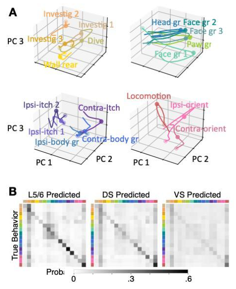
Fig 4: A) Neural trajectories aligned to spontaneous behaviors have predictable structure and occupy different parts of neural state space. B) Action type can be decoded in Motor Cortex Layer 5/6 and Dorsal Striatum, but predictions are optimal in cortex. Ventral Striatum provided as a ‘non-motoric’ reference.
Psychiatric and Clinical Dimensions
The ability to readily capture and accurately quantify previously inaccessible behaviors has proven to be a powerful and sought after tool across a range of disease states. Together with collaborators in the Pittsburgh-area and internationally, we have quantified brain-behavior dysfunction in several disease states. First, we helped uncover several behavioral changes in a recently developed mouse model of Huntington’s disease, showing different manifestations of the disease across sex, age, and background strain (Koch et al., BMC Biology). In another study, we revisited our focus on OCD, helping to reveal how overexpression of a specific glutamate transporter in the forebrain increases susceptibility to amphetamine induced OCD-like behaviors (Kopelman et al., eNeuro). In each case, our collaborators and lab members were able to connect basic biological function of cortico-striatal circuits to complex behavioral phenotypes.
Our expertise has made for valued collaboration, particularly in pain research where new behavioral insights can produce large advances in translational understanding. With colleagues at Columbia and Tufts, we used B-SOiD alongside other computational approaches to map the progression of pain states in mice — uncovering distinct strings of spontaneous behavioral syllables representing pain, pain relief, and recovery in freely moving animals (Bohic et al., Neuron). A separate study with close collaborators at UPenn discovered that a specific population of anterior cingulate cortex neurons encodes spontaneous pain-related behaviors and is selectively modulated by morphine, an effective but addictive analgesic or pain reliever. With the multi-dimensional behavioral structure we provided, the team was able to show that morphine reduces affective-motivational behaviors without altering sensory detection or reflexive responses. Using this as a baseline, we went on to demonstrate that a novel chemogenetic gene therapy developed by our co-authors mimics morphine analgesia at the behavioral level, but without the associated risks of addiction (Oswell et al., Nature; Fig 5). To do so, we created an all-in-one physical hardware and pain-specific B-SOiD software package, LUPE, enabling the community to access complex behavioral phenotypes at one-tenth of the price of recently released commercial options.
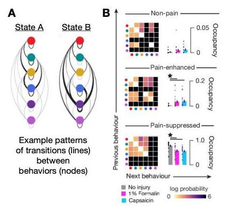
Fig 5: A) Example of structured transition probabilities between B-SOiD-identified behaviors. B) We found that transition state patterns could readily identify pain condition.
Future Directions
Realtime behavior detection to trigger closed-loop manipulations. While offline comparison of neural and behavioral dynamics can be informative, my career has taught me the power of closed-loop manipulations in understanding the coupling of brain to behavior. Through the application of new advances in computer vision, we are side-stepping the computational sand traps that slow down behavior classifiers. Soon, we will be able to deliver optogenetic stimulation precisely (<10ms latency) when an animal enters a specific behavioral state. We will then apply this tool in adaptive experimental designs – similar to those in Fig 1 that have been central to our research program – to more deeply understand how multi-area neural dynamics shape and orchestrate behavior.
Expansion of disease specific behavior analysis platforms. Given our successes in characterizing the behavioral phenotypes associated with pain and analgesia, we are expanding into two more disease states: autism spectrum disorder and systemic infection (“feeling ill”). While the former is in its initial stages with fellow department members Zheng Kuang and Kate Hong, the latter will be submitted as an R01 proposal this summer with co-PI Jessica Osterhout. This work builds off of a standing collaboration and Osterhout’s recent Nature study that discovered a population of previously uncharacterized hypothalamic neurons responsible for the cavalcade of sickness symptoms and behavioral changes that come about as a result of infection. Our group will lead the proposal, building a multi-dimensional behavioral model of sickness and health that for the first time will also include physiological signals (respiration, temperature) – with the end goal of predicting infection severity and benchmarking new therapeutic solutions.
Potentiating our efforts through institutional implementation. To multiply the impact of our work, we are actively in talks with the NIH to install our LUPE platform as a central pillar of NIDA’s Neural Ensembles & Used Substances (NExUS) ‘Collaboratory’. We are also working with the NIH to establish A/B-SOiD as an analytical cornerstone of the Brain Behavior Quantification and Synchronization (BBQS) Consortium and its subsidiary members, including EMBER, DANDI, and Neurodata Without Borders.