Our lab takes an interdiscplinary approach to developmental biology. We incorporate time-lapse microscopy, fluorescent probe development and proteomics to address longstanding questions about embryo development.
Proteomics
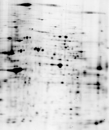
Proteomics is the analysis of all proteins in a cell, tissue, or organism. We have developed new methods for proteome analysis. Cells constantly have to adapt to environmental changes and mutational changes. This adaptation is carried out by changes in protein expression and protein modification. We have developed a powerful new method to rapidly identify protein differences between cells. This method, called Difference Gel Electrophoresis (DIGE), involves fluorescently tagging the proteins of different cell extracts with different color fluorescent dyes. The differently colored proteins are mixed and run on the same two-dimensional electrophoresis gel. We then take two pictures of the gel, selecting the wavelength specific for each color. Comparing the images allows one to quickly identify which proteins differ between the samples. We have used this method to identify protein changes during Drosophila embryo development, in Drosophila behavioral mutants, in cancer cells, in mutant yeast and a wide range of organisms and conditions.
Shown here is a two-frame, looping movie of a 2D-DIGE gel comparing wild-type mouse cells to cells from a knock-out mouse. Horizontal shifts indicate changes in post-translation modification.
Time-lapse Microscopy and Fluorescent Probe Development
Multicellular organisms develop by transforming a mass of undifferentiated cells into intricately organized groups of cells that coalesce to form functional structures. This process is known as pattern formation. We are interested in what happens when pattern formation is altered: How does the developing embryo respond when too few or too many cells are segregated to form a particular structure? Drosophila embryos have a tremendous capacity to repair mispatterned tissues. Using genetically altered embryos, we can study the cell biology of pattern repair in a wide variety of ways. We have taken an interdisciplinary approach to understanding pattern repair, from genetics and molecular biology to high-end microscopy and computerized image analysis to the design and synthesis of new chemical reagents. We have developed methods to follow cell division, cell shape, gene expression and cell death by time-lapse microscopy. We have also developed a method for turning selected genes on in specific cells with light, called photoactivated gene expression. This allows us to alter the behavior of single cells,or patches of cells, and determine how the cells respond. We have discovered that cell death plays a major role in repairing these induced pattern defects. We are currently investigating how this pattern-repair-induced cell death is controlled.