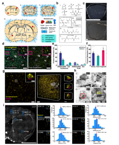
Our laboratory is pioneering innovative approaches to address the challenges of understanding complex biological systems. These are finely tuned ensembles of nanoscale building blocks like proteins, nucleic acids, lipids, and carbohydrates. We focus on developing technologies that allow large-scale visualization of these biological samples with unprecedented nanoscale precision. Central to our efforts is the principle of expansion microscopy (ExM). This groundbreaking technique physically expands the sample, offering a novel alternative to traditional magnification methods that rely on lenses. Building on this, we have developed Expansion Pathology (ExPath), an advanced form of ExM tailored for detailed nanoscale imaging of various human tissue samples, including those prepared with traditional methods like paraffin-embedding and hematoxylin and eosin staining. ExPath achieves this by embedding specimens in a swellable polymer, equalizing their mechanical properties, and then expanding them up to fivefold, thus achieving a resolution of approximately 60 nm. This technique combines the throughput of a diffraction-limited microscope with the resolution of a super-resolution microscope, allowing for diagnosing diseases that traditionally required electron microscopy. Further extending our research, we have also introduced next-generation ExM technologies such as Magnify, MAGNIFIER, and microMagnify, pushing the resolution boundaries to approximately 15 nm across a wide array of biological samples. These cutting-edge technologies hold immense potential for revealing intricate details of complex systems like the brain, cancer, and infectious diseases, opening new avenues for understanding and treating these conditions.
Selected Publications
- Cheng, C. Stefani, T. Skillman, A. Klimas, A. Lee, E. DiBernardo, K. Brown, T. Milman, Y. Wang, B. Gallagher, K. Lagree, B. Jena, J. Pulido, A. Mitchell, S. Filler, L. Hiller, A. Lacy-Hulbert, Y. Zhao*, “MicroMagnify: a multiplexed expansion microscopy method for pathogens and infected tissues“,Advanced Science, 2023, 2302249. [Editors’ choice]
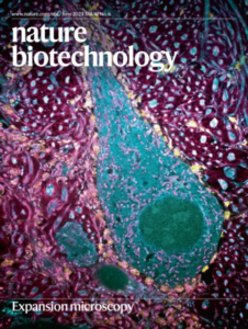
- [Cover Article] A. Klimas, B. Gallagher, P. Wijesekara, S. Fekir, E. F. DiBernardo, Z. Cheng, D. Stolz, F. Cambi, S. Watkins, S. L. Brody, A. Horani, A. L. Barth, C. Moore, X. Ren, Y. Zhao*, “Magnify is a universal molecular anchoring strategy for expansion microscopy”.Nature Biotechnology 41, pages858–869 (2023). Full text is available here.
- L. Shi, A. Klimas, B. Gallagher, Z. Cheng, F. Fu, P. Wijesekara, Y. Miao, X. Ren, Y Zhao* (co-corresponding author), W Min*, “Super-resolution vibrational imaging using expansion stimulated Raman scattering microscopy”, Advanced Science,2022, 2200315.
- Y. Zhao (equal contribution), O. Bucur (equal contribution), H. Irshad, F. Chen, A. Weins, A. L. Stancu, E – Y. Oh, M. DiStasio, V. Torous, B. Glass, I. E. Stillman, S. J. Schnitt, A. H. Beck*, E. S. Boyden*, ‘Nanoscale imaging of clinical specimens using pathology-optimized expansion microscopy.’ Nature Biotechnology, 2017; DOI: 10.1038/nbt.3892 [See also MIT News and BIDMC News]
- P. W. Tillberg, F. Chen, K.D. Piatkevich, Y. Zhao, C.-C. Yu, B. P. English, L. Gao, A. Martorell, H.-J. Suk, F. Yoshida, E. M. DeGennaro, D. H.Roossien, G.. Gong, U. Seneviratne, S. R. Tannenbaum, R. Desimone, D. Cai, E. S. Boyden*, ‘Protein-retention expansion microscopy of cells and tissues labeled using standard fluorescent proteins and antibodies’, Nature Biotechnology, 2016, 34 (9), 987–992. [Cover article in September 2016 issue of Nature Biotechnology (volume 34, No. 9).]